

Fractured Ti-Base
Diagnosing and Managing a Dental Implant Crown with a Horizontally Fractured Ti-Base: A Specialist’s Approach
Dental implants are a reliable and long-lasting solution for replacing missing teeth. However, complications can arise, such as a horizontally fractured Ti-base (titanium base) beneath the implant crown. This article details how a dental implant specialist would diagnose and manage this issue, ensuring the patient receives the best possible care.
Step 1: Diagnosis
The process begins with a thorough examination to confirm the problem and determine the extent of the damage.
- Patient Symptoms:
- The patient may report a loose crown, discomfort while chewing, or a visible crack in the crown.
- In some cases, there may be no symptoms, and the fracture is discovered during a routine check-up.
- Clinical Examination:
- The specialist visually inspects the implant crown and surrounding tissues for signs of damage or inflammation.
- The crown is gently tested for mobility or movement, which may indicate a fractured Ti-base.
- Radiographic Imaging:
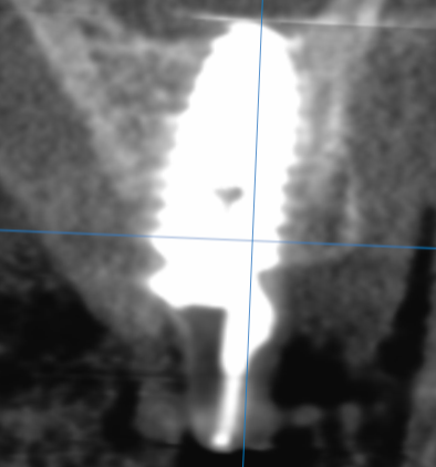
- A periapical X-ray or cone beam computed tomography (CBCT) scan is taken to assess the implant and Ti-base.
- The imaging helps confirm the horizontal fracture and evaluate the integrity of the implant fixture and surrounding bone.
- Removal of the Crown:
- The crown is carefully removed to inspect the Ti-base directly. A fractured Ti-base will typically show a visible horizontal crack.
Step 2: Management and Treatment
Once the diagnosis is confirmed, the specialist develops a treatment plan tailored to the patient’s needs.
Option 1: Replacement of the Ti-Base and Crown
- If the implant fixture is intact and the surrounding bone is healthy, the fractured Ti-base can be replaced.
- Procedure:
- The old crown and Ti-base are removed.
- A new Ti-base is selected and attached to the implant fixture.
- A new crown is fabricated using digital scanning and CAD/CAM technology for a precise fit.
- The new crown is cemented or screwed onto the Ti-base.
Option 2: Implant Removal and Replacement
- If the implant fixture is also damaged or the bone support is compromised, the entire implant may need to be replaced.
- Procedure:
- The fractured Ti-base, crown, and implant fixture are removed.
- The site is allowed to heal, and bone grafting may be performed if necessary.
- A new implant is placed once the site is ready, followed by a new Ti-base and crown.
Step 3: Prevention and Maintenance
To prevent future complications, the specialist provides the patient with guidance on maintaining their implant and crown:
- Oral Hygiene: Brush and floss regularly to keep the implant and surrounding tissues healthy.
- Avoid Excessive Force: Do not use the implant crown to bite hard objects or open packages.
- Regular Check-Ups: Visit the dentist every 6 months for professional cleanings and examinations.
- Night Guards: If the patient grinds their teeth, a custom night guard can protect the implant and crown.
How Digital Dentistry Enhances the Process
- Digital Scanning: Intraoral scanners create precise 3D models of the implant site, ensuring accurate fabrication of the new Ti-base and crown.
- CAD/CAM Technology: Custom crowns are designed and milled with high precision, reducing treatment time and improving fit.
- CBCT Imaging: Provides detailed 3D views of the implant and surrounding structures, aiding in diagnosis and treatment planning.
Final Thoughts
A horizontally fractured Ti-base is a rare but manageable complication in dental implant treatment. With advanced diagnostic tools like CBCT imaging and modern techniques like CAD/CAM technology, a dental implant specialist can effectively diagnose and treat the issue, restoring the patient’s smile and function.
By addressing the issue promptly and using state-of-the-art technology, the specialist ensures the patient achieves a long-lasting and successful outcome.
If you are having problems with your dental implant crown, Contact Us today to see how we can help you.






Recent Comments